Home
/ Neck Muscle Diagram Back : Back And Neck Muscles Graph Diagram / Muscles, connected to bones or internal organs and blood vessels, are in charge for movement.
Neck Muscle Diagram Back : Back And Neck Muscles Graph Diagram / Muscles, connected to bones or internal organs and blood vessels, are in charge for movement.
Neck Muscle Diagram Back : Back And Neck Muscles Graph Diagram / Muscles, connected to bones or internal organs and blood vessels, are in charge for movement.. Learn this topic fast with head and neck muscle anatomy reference charts. It runs superolaterally from the back to top. Neck muscles are divided into separate groups according to their origin and topographic features (by neck areas). Posted on december 4, 2018december 3, 2018. Axial muscles of the head neck and back anatomy and, imテ genes fotos de stock y vectores sobre muscle chart, , back of neck anatomy diagram vector illustration of neck muscles anatomy.
Muscles, connected to bones or internal organs and blood vessels, are in charge for movement. This is a table of skeletal muscles of the human anatomy. The back's muscles start at the top of the back (named the cervical vertebrae) and go to the tailbone (also named the coccyx). Equally important is the erector spinae muscles. Musculoskeletal, shoulder & back neck muscles.
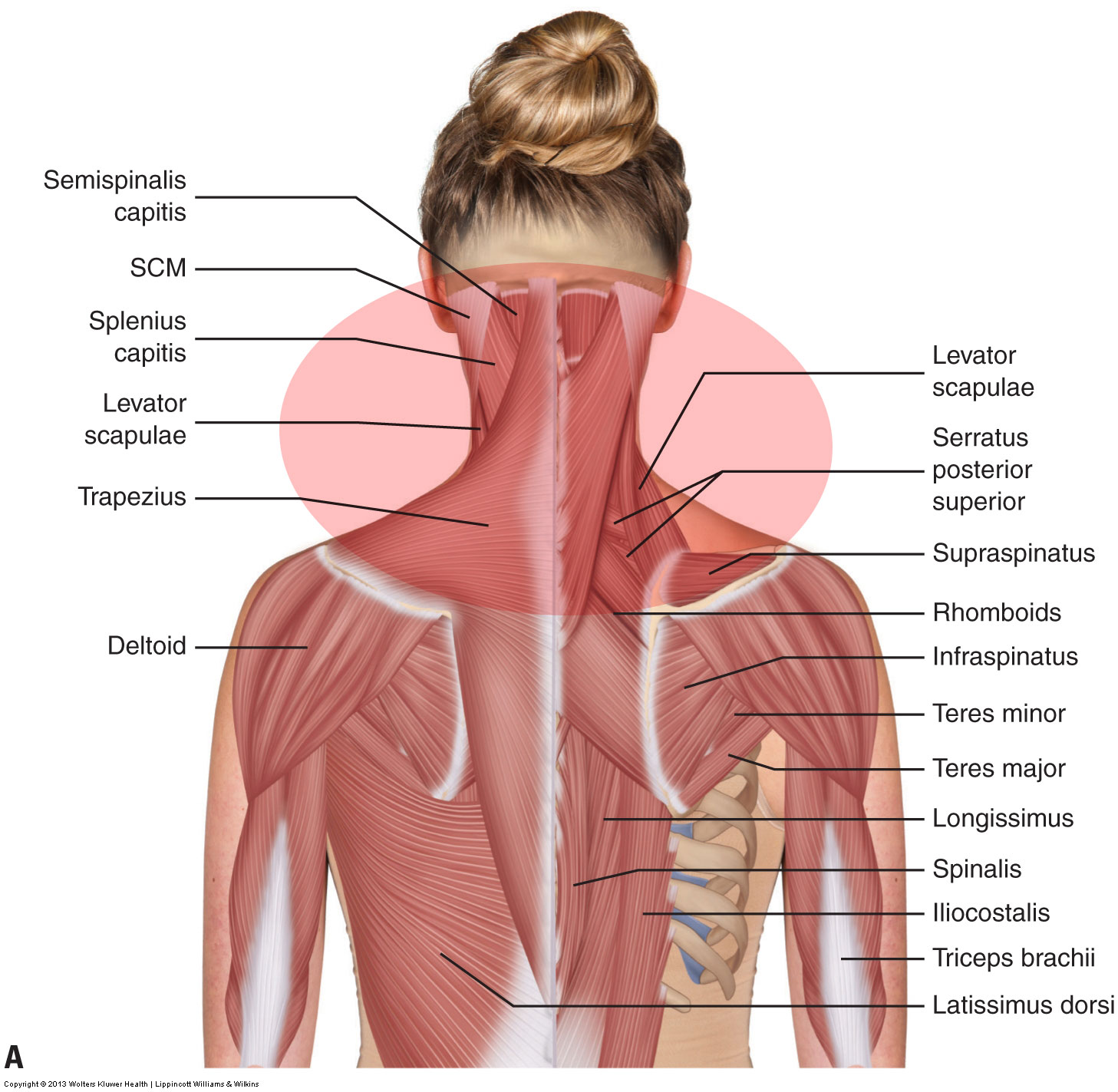
Labeled Anatomy Chart Of Neck And Back Muscles On White Background Stock Photo Download Image Now Istock from media.istockphoto.com Working in pairs on the left and. Neck muscles help support the cervical spine and contribute to movements of the head, neck, upper back, and shoulders. The trapezius muscle actually considered to be just as much of a muscle related to the back as it is the neck. Advertisements help pay for this website. Muscle anatomy body anatomy deadlift muscles worked. Discover ideas about muscle diagram. Neck muscle diagram back : They are divided into three groups, as shown below.
First rule of doing superheroes, learn the muscles!
The back's muscles start at the top of the back (named the cervical vertebrae) and go to the tailbone (also named the coccyx). Advertisements help pay for this website. Head and neck muscles diagram so many muscles that cause migraines arm neck shoulders and back templates. Platysma, sternocleidomastoid, splenius capitis, levator scapulae, scalene muscles, omohyoid, trapezius, sternothyroid, thyrohyoid, omohyoid, sternohyoid. The trapezius muscle actually considered to be just as much of a muscle related to the back as it is the neck. They move the head in every direction, pulling the skull and jaw towards the shoulders, spine, and scapula. Cadillac bose amp wiring diagram. Their main function is contractibility. The neck muscles are specifically designed to either allow for neck movement or to provide structural support for the head. When we think of back muscles, latissimus dorsi (lats) comes to mind. The suprahyoid muscles are a group of neck muscles located above the hyoid bone and all elevate this bone, while the infrahyoid muscles are situated several other muscles of the back also extend up to the neck region and are partly connected with the cervical part of the vertebral column, including. Thank you for your support. The muscles of the neck:
Neck muscle diagram back : Almost every movement in the body is the outcome of muscle contraction. The drawings here present idealized versions of male and female torsos. Muscles, connected to bones or internal organs and blood vessels, are in charge for movement. The trapezius muscle is a large surface muscle that spans from the base of the skull down the cervical spine and into the lower thoracic spine (mid back), as well as out to the.
Pin On Dental School from i.pinimg.com The deep back muscles lie immediately adjacent to the vertebral column and ribs. Neck muscle of back deep. The anterior and middle scalenes originate from the transverse processes of certain cervical. Peter wu here is a presentation with diagrams of the muscles in the neck. Head and neck anatomical chart. Neck neck muscle anatomy muscle diagram inspirational medical. The three scalene muscles are found forming the floor of the posterior triangle. The trapezius muscle actually considered to be just as much of a muscle related to the back as it is the neck.
It runs superolaterally from the back to top.
Discover ideas about muscle diagram. This thin muscle tenses the skin of the neck. Luckily you've found this page to help you. The major muscle of the back of the neck, the trapezius, is involved in movements of the scapula and is dealt with in the next section, on the muscles of muscle diagram. Upper back and neck muscles | the erector spinae muscles work together to keep the spine erect as. Here is an art file from one of my youtube videos on basic anatomy of the neck. Below you'll see diagrams along with the names of the back muscles that may be the cause of your these are strong, large muscles are located on either side of the neck. Back muscle diagram human body, back muscle diagram pain, back muscle groups diagram posterior, neck muscle anatomy ultrasound, neck muscles anatomy radiology, human muscles, neck muscle anatomy images, neck muscle anatomy pictures. The back muscles can be three types. Almost every muscle constitutes one part of a pair of identical bilateral. The anterior and middle scalenes originate from the transverse processes of certain cervical. The trapezius muscle is a large surface muscle that spans from the base of the skull down the cervical spine and into the lower thoracic spine (mid back), as well as out to the. They move the head in every direction, pulling the skull and jaw towards the shoulders, spine, and scapula.
The anterior and middle scalenes originate from the transverse processes of certain cervical. First rule of doing superheroes, learn the muscles! Best back muscles training exercises / equally important is the erector spinae muscles. 12 photos of the back muscle chart. Back muscle diagram human body, back muscle diagram pain, back muscle groups diagram posterior, neck muscle anatomy ultrasound, neck muscles anatomy radiology, human muscles, neck muscle anatomy images, neck muscle anatomy pictures.
What Are The Causes Of Muscle Spasming In The Neck from learnmuscles.com Working in pairs on the left and. Luckily you've found this page to help you. The back muscles can be three types. Get a britannica premium subscription and gain access to exclusive content. There are around 650 skeletal muscles within the typical human body. The anterior and middle scalenes originate from the transverse processes of certain cervical. Er diagram to relational schema. Human muscle system functions diagram facts britannica.
Er diagram to relational schema.
Head and neck anatomical chart. Musculoskeletal, shoulder & back neck muscles. Splenius capitis is one of the deep back muscles that is associated with rotating and extending the head and neck. Superficial muscles are the muscles closest to the skin surface and can usually be seen while a body is performing actions. Intermediate back muscles and c. At least that was my approach back in the day. Since the all the back muscles originate in embryo (fetus). The neck muscles are specifically designed to either allow for neck movement or to provide structural support for the head. The anterior and middle scalenes originate from the transverse processes of certain cervical. Distinguish the muscles that developed on the basis of the first (mandibular) and second (hyoid) visceral, gill arches, and muscles that developed from the ventral divisions of the myotomes. Platysma, sternocleidomastoid, splenius capitis, levator scapulae, scalene muscles, omohyoid, trapezius, sternothyroid, thyrohyoid, omohyoid, sternohyoid. Almost every movement in the body is the outcome of muscle contraction. Automatic on off switch for water pump circuit diagram.
Axial muscles of the head neck and back anatomy and, imテ genes fotos de stock y vectores sobre muscle chart, , back of neck anatomy diagram vector illustration of neck muscles anatomy neck muscle diagram. The anterior and middle scalenes originate from the transverse processes of certain cervical.